CCBIR Virtual Symposium (1/30/2025)
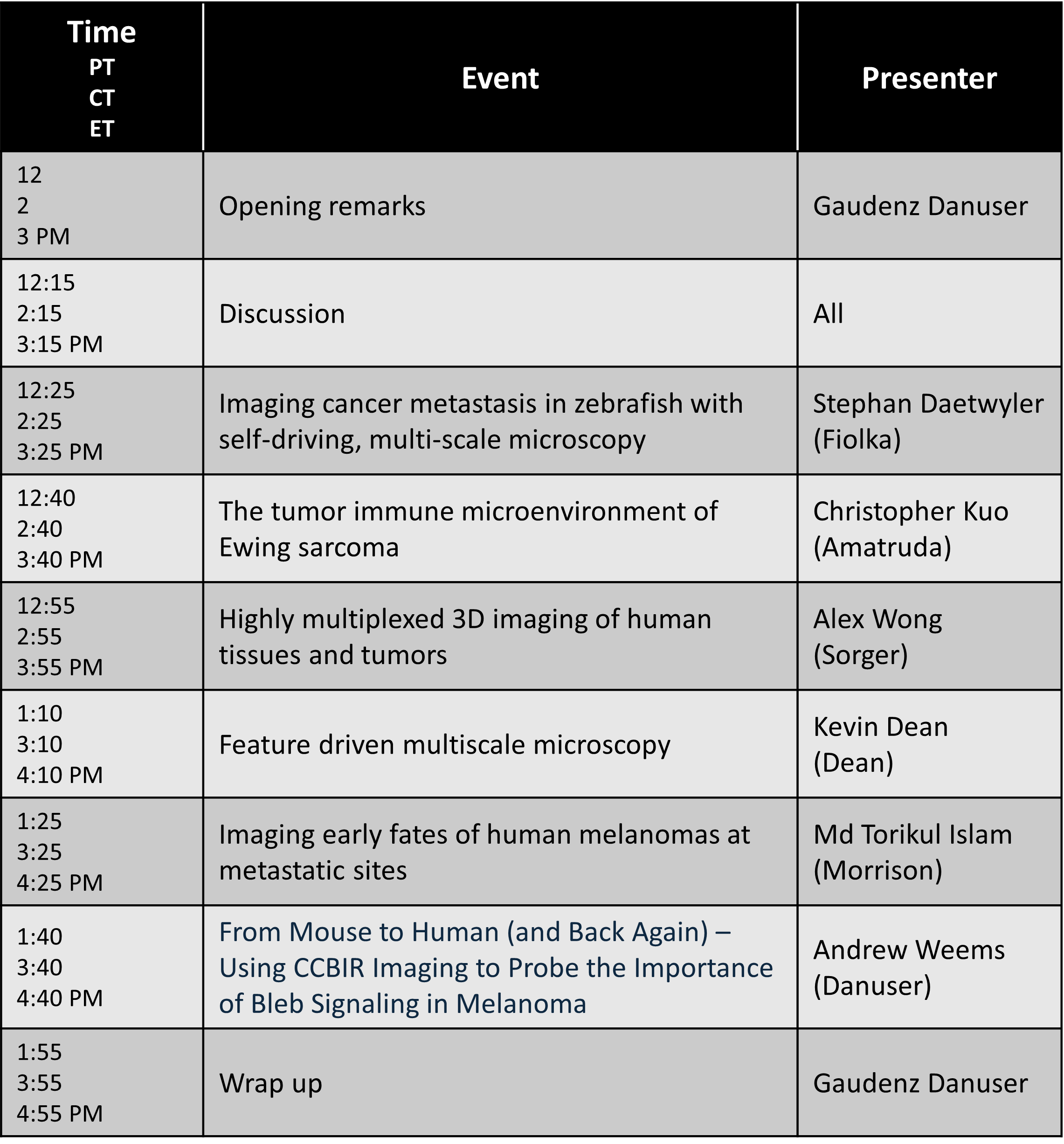
Imaging cancer metastasis in zebrafish with self-driving, multi-scale microscopy
Stephan Daetwyler, PhD (postdoc - Fiolka Lab, UT Southwestern)
Zebrafish xenografts and cancer models provide powerful model systems to study the dynamic, multi-factorial processes of cancer metastasis in a near-physiological environment in vivo. To observe and quantify subcellular behaviors of cancer cells alongside entire organisms over days of imaging, we introduce a versatile self-driving, multi-resolution light-sheet microscope platform. We demonstrate its power by imaging cancer invasion, cancer spreading and immune-cancer cells interactions that lead to either immune evasion or phagocytosis.
The tumor immune microenvironment of Ewing sarcoma
Christopher Kuo, MD (attending physician - Amatruda Lab, Children’s Hospital Los Angeles)
To date, there is a lack of understanding of the tumor microenvironment (TME) of Ewing sarcoma (EwS). By performing spatially resolved transcriptomics to primary treatment-naïve EwS samples, we discovered greater stromal enrichment in localized EwS tumors compared to metastatic EwS tumors. Through spatial ligand-receptor analysis, we show that the stromal enriched regions harbor unique extracellular matrix related cytokines, immune recruitment and proinflammatory microenvironmental signals, implying EwS stroma may play an anti-tumorigenic role by acting as an immune recruitment center.
Highly Multiplexed 3D Imaging of Human Tissues and Tumors
Alex Wong, PhD (postdoc - Sorger Lab, Harvard Medical School)
By adapting light-sheet microscopy for 3D cyclical imaging and refining human tissue chemistries for optical clearing and staining, we investigate neuro-immune interactions and analyze morphological differences between adjacent normal and colorectal adenocarcinoma tissues.
Feature Driven Multiscale Microscopy
Kevin Dean, PhD (Assistant Professor, UT Southwestern)
Multiscale Cleared Tissue Axially Swept Light-Sheet Microscopy (MCT-ASLM) seamlessly integrates macroscale and nanoscale imaging modules and enables feature-driven, multi-resolution imaging of chemically cleared tissues. By automating the identification and interrogation of biological features, such as glomeruli in kidneys and metastatic colonies, MCT-ASLM bridges spatial scales, providing a transformative approach for linking subcellular structures to tissue-wide architectures.
Imaging Early Fates of Human Melanomas at Metastatic Sites
Md Torikul Islam, PhD (postdoc - Morrison Lab, Children’s Research Institute, UT Southwestern)
Human melanomas demonstrate remarkable heterogeneity in their ability to metastasize to distant sites. Using xenograft assays, we aim to image the early stages of spontaneous metastasis to understand the mechanisms that limit metastasis and determine organotropism. We found that only a small subset of cells gives rise to the majority of metastatic disease burden, and metastatic signals are detected in the lungs before other organs.
From Mouse to Human (and Back Again) -- Using CCBIR Imaging to Probe the Importance of Bleb Signaling in Melanoma
Andrew Weems, PhD (Assistant Professor, UT Austin)
Our recent work has led to the discovery of Bleb Signaling -- a novel cytomorphic signaling pathway upon which melanoma cells rely for survival. Though compelling, our findings have thus far been limited to observations and experiments done in vitro using melanoma cell lines. Through intellectual and technical collaborations driven by the CCBIR program, we now present results demonstrating the occurrence and importance of this pathway in developing cancer systems in vivo, as well as the therapeutic potential of targeting the pathway in the clinic.
Our team members attended the 2023 CCBIR Annual Investigators Meeting June 6-8!
Talks
Reto Fiolka - Multi-scale Imaging of Cancer Metastasis in Zebrafish Xenograft Models
Zach Marin - Imaging metastatic proliferation in situ
Felix Zhou - Towards complete quantitative subcellular mapping of single cells and molecular signals in 3D tissue microenvironments
Dagan Segal - In vivo 3D profiling of site-specific human cancer cell morphotypes in zebrafish
Md Torikul Islam - Determining melanoma cell fates in metastatic sites
Andrew Weems - Bleb Signaling in Cancer Cell Survival and Disease Progression
Posters
Hazel Borges - Optimize imaging of fluorescently labeled and chemical cleared tissues
Bo-Jui Chang - Light-sheet fluorescence microscope for cancer research
Md Torikul Islam - Determining melanoma cell fates in metastatic sites
Tada Isogai - Anchorage-independent cell proliferation mediated by F-actin bundling
Jinlong Lin - Nanoscale subcellular biology with Expansion Axially Swept Light-Sheet Microscopy
People’s Choice Award - Staff Scientist
Dagan Segal - Caveolin-1 organization and function drive functional plasticity in Ewing Sarcoma
Felix Zhou - Towards complete quantitative subcellular mapping of single cells and molecular signals in 3D tissue microenvironments
Conor McFadden - Towards high-throughput imaging of cancer metastatic niches in vivo
Stephan Daetwyler - Self-Driving, multi-scale microscopy capturing cancer cell dynamics with high resolution in vitro and in situ
People’s Choice Award - PostDoc
Kevin Dean - Imaging the Metastatic Cascade with Axially Swept Light-Sheet Microscopy
From the Sorger Lab at Harvard Medical School!
Unlike standard immunofluorescence, cyclic immunofluorescence can identify more than 60 proteins on the same sample. We are using cyclic immunofluorescence on patient tissues to understand how diseases such as cancer begin. In this video, we found tumor cells stained by MART1 (green) and alot of CD8-positive cytotoxic immune T cells (yellow) in a melanoma sample. However, the immune cells also stained with LAG3 (red dots) meaning that they are less effective at combating cancer. By combining cyclic immunofluorescence with 3D imaging, we aim to uncover additional features of cancer cells, such as their volume, shape, and location, that will follow the progression of disease more accurately.
3D imaging of a human melanoma sample with cyclic immunofluorescence
From the Dean lab at UT Southwestern!
Depicted here is a 3D image of a lung where all of the nuclei have been segmented. This allows us to count every cell in the tissue, including those that are metastatic.